
Table of Contents
A pure-tone audiogram can tell us how softly a person hears across a range of frequencies. But one set of thresholds is not enough to tell us where a hearing difficulty begins.
That is where bone-conduction testing earns its place.
Air-conduction testing sends sound through the outer ear, eardrum and middle ear before it reaches the cochlea. Bone-conduction testing takes a different route. A small vibrator placed behind the ear sends mechanical vibration through the skull to stimulate the cochlea, largely bypassing the outer and middle ear.
Comparing those two routes can reveal whether reduced hearing sensitivity is mainly associated with sound transmission through the outer or middle ear, with the inner ear, or with a combination of both.
In practice, interpretation requires care. Bone conduction is not a shortcut to a diagnosis, and an apparent difference between air and bone thresholds is not automatically proof of middle-ear disease. The result has to be read alongside otoscopy, tympanometry, the hearing history and the rest of the audiogram.
Air and bone conduction ask different questions
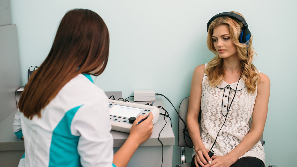
During air-conduction audiometry, tones are presented through headphones or insert earphones. Sound follows the same broad route as everyday sound: through the ear canal, across the eardrum and middle-ear structures, and onwards to the cochlea.
If the threshold is elevated, we know hearing sensitivity is reduced at that frequency. Air conduction alone does not tell us which part of the auditory pathway is responsible.
Bone conduction helps separate the problem.
The vibrator is usually placed on the mastoid bone behind the ear. Its vibration reaches the inner ear through the skull, reducing the influence of the ear canal, eardrum and middle-ear system on the measured threshold.
If bone-conduction thresholds are substantially better than air-conduction thresholds, that suggests the outer or middle ear is contributing to the loss measured through headphones.
If air and bone thresholds are elevated to a similar degree, the pattern is more consistent with sensorineural hearing loss. If bone thresholds are elevated but air thresholds are poorer still, there may be both sensorineural and conductive components: a mixed hearing loss.
These patterns classify hearing loss; they do not identify the exact medical cause.
A conductive component could relate to several conditions, from earwax or middle-ear fluid to eardrum or ossicular problems. Examination and, when appropriate, tympanometry or ENT assessment help explain why.
The air-bone gap is useful, but it is not a diagnosis
The difference between air- and bone-conduction thresholds at the same frequency is known as the air-bone gap.
When that gap is clinically meaningful and repeatable, it can indicate a conductive component. But the number needs context.
Audiometric thresholds have normal test-retest variability, so small differences can occur without representing pathology. Professional protocols also use specific criteria to decide when masking is required and when a gap should influence classification.
Recent evidence adds another caution. False air-bone gaps can occur even in ears with confirmed sensorineural hearing loss, particularly at certain frequencies. Technical limitations of bone vibrators, calibration effects and vibrotactile responses can all contribute.
This matters at very low frequencies, where a person may sometimes feel vibration rather than hear it, and at higher bone-conduction frequencies, where transducer and calibration limitations become more significant.
For that reason, an audiologist should not look at one isolated gap and immediately label the loss conductive.
The pattern across frequencies matters. So do otoscopic findings, middle-ear measurements, symptoms and previous results. A convincing conductive pattern is usually supported by more than a single point on the graph.
Why masking is sometimes essential
Bone-conduction testing introduces a problem that is easy to miss if you only look at the equipment.
When the skull vibrates, both cochleae can potentially detect that vibration. Placing the bone vibrator behind the right ear does not guarantee that the right cochlea is the one responding.
That becomes important when the two ears hear differently.
Suppose we are trying to establish the bone-conduction threshold of the poorer ear. Without additional steps, the better cochlea may respond first. The resulting threshold could then be wrongly attributed to the ear we intended to test.
Masking is how audiologists separate the ears.
Narrow-band noise is presented to the non-test ear so that it cannot respond to the test tone. The bone-conduction threshold can then be established more confidently for the ear under investigation.
Masking is also used in air-conduction testing when there is enough asymmetry for a strong test signal to cross the skull and be detected by the opposite ear.
The procedure can feel repetitive because the audiologist may need to adjust both test tone and masking noise several times. That repetition is what allows an apparently simple threshold to become ear-specific.
Asking the patient which ear they heard the bone-conducted tone in is not a reliable substitute; people often cannot localise the vibration accurately.
Bone conduction has technical limits of its own
Bone-conduction testing is indispensable, but it is not as unrestricted as air-conduction audiometry.
The vibrator has a narrower useful frequency and output range than conventional earphones. Professional guidance therefore advises caution where distortion, calibration limitations or tactile responses can make results less reliable.
Placement matters too. The vibrator must sit correctly on the mastoid, with appropriate headband pressure and without resting on hair or touching the pinna. Changes in coupling can alter the measured response.
At low frequencies and higher presentation levels, vibration may be felt through the skin or skull. A patient who responds because they feel the stimulus rather than hear it can produce an apparent threshold that does not represent auditory sensitivity.
There are also situations in which the bone vibrator cannot produce enough output to establish a true threshold. The audiogram must then show that no response was obtained at the equipment limit rather than imply that the exact threshold is known.
A 2026 study of 1,000 ears provides a useful reminder. Clinically significant false air-bone gaps were particularly common at 250 Hz and also occurred at 4000 Hz in ears classified as sensorineural hearing loss. The finding reinforces the need to cross-check an apparent gap against the wider clinical picture.
The value is in the comparison, not the headband
Patients sometimes assume that bone-conduction testing is an “extra” hearing test used only when the first test has gone wrong.
It is better understood as a second route to the same destination.
Air conduction tells us how softly sound can be detected through the whole peripheral hearing pathway. Bone conduction reduces the influence of the outer and middle ear. Comparing the two helps classify the pattern of hearing loss and decide what information is still missing.
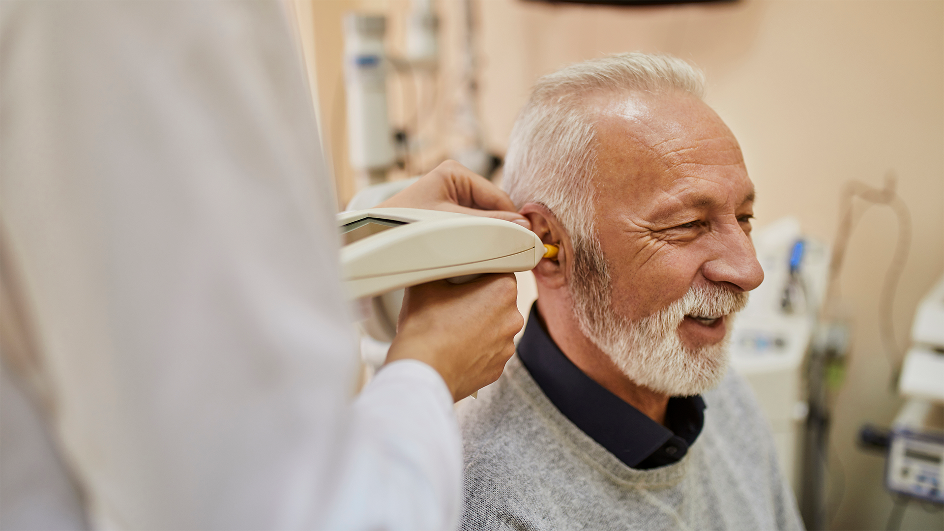
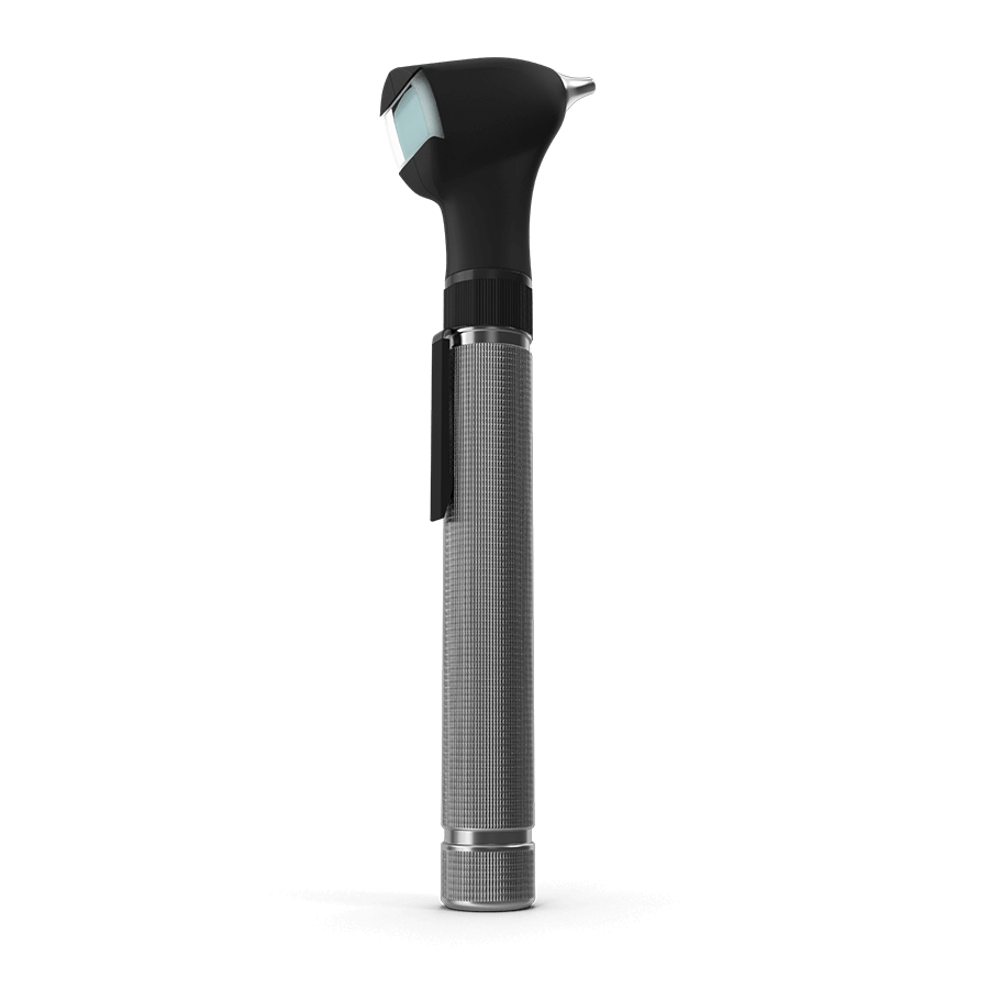
At AudioCare, that comparison can sit alongside video otoscopy, tympanometry, speech audiometry and other tests selected according to the person’s history and symptoms.
A patient with an air-bone gap and abnormal middle-ear findings requires a different interpretation from someone with elevated air and bone thresholds that closely match. An asymmetric result may require masking or further investigation. A result that does not fit the clinical history may need to be repeated or cross-checked rather than accepted at face value.
The point is not to perform every available test on every patient. It is to know which question needs answering.
Bone-conduction testing answers a particularly important one: is reduced hearing sensitivity being caused, at least in part, by the route sound takes through the outer and middle ear, or does the pattern remain reduced even when those structures are largely bypassed?
That distinction can change the type of hearing loss recorded on the audiogram, the need for additional testing and whether medical or ENT referral is appropriate.
The headband itself is simple.
What it adds to the audiogram is not.
References
- https://www.thebsa.org.uk/wp-content/uploads/2023/10/OD104-32-Recommended-Procedure-Pure-Tone-Audiometry-August-2018-FINAL-1.pdf
- https://baaudiology.org/updates-to-bsa-documents/
- https://www.asha.org/practice-portal/clinical-topics/hearing-loss/
- https://www.asha.org/policy/GL2005-00014/
- https://doi.org/10.1044/2026_AJA-26-00029
- https://audiocare.pt/pure-tone-speech-audiometry/